

Quick answer: Retatrutide (LY3437943) is a synthetic, 39-residue peptide. Its structure includes noncanonical amino-acid substitutions and a C20 fatty-diacid side chain attached at Lys17 through a linker. These features are part of the molecule’s design; a schematic or sequence record is not evidence that a separately marketed sample has the same identity or quality.
This article addresses the searches “retatrutide structure” and “retatrutide chemical structure” by explaining what a sequence record describes, how the principal modifications are represented, and why different database entries may show different salt forms. It is a research reference, not a synthesis, handling, or personal-use guide.
What is the retatrutide structure?
Retatrutide is also known by the development code LY3437943. Published structural and pharmacology research describes it as a single peptide with activity at GIP, GLP-1, and glucagon receptors. The molecule is commonly described as a 39-residue peptide with a lipid side-chain modification.
“Structure” can refer to several related records: the order of amino-acid residues, nonstandard residue chemistry, a covalently attached side chain, a two-dimensional structure drawing, or a three-dimensional conformation. These are not interchangeable. A simple one-letter sequence may omit noncanonical residues, linkers, terminal chemistry, or salt form.
Backbone versus side-chain modification
The peptide backbone is the linked residue chain. In retatrutide, a fatty-diacid moiety is attached to a lysine side chain through a linker. The lipid appendage is covalently connected to the peptide; it is not a second active peptide or a separate ingredient in the sequence.
How is the retatrutide sequence organized?
Public chemical references describe a 39-residue sequence derived from incretin-peptide design, with several substitutions that are not standard one-letter amino-acid codes. An unmodified alphabetic string can therefore be misleading if it replaces these residues with approximate symbols or leaves them out entirely.
Noncanonical residues reported in the structure
- Aib at positions 2 and 20: Aib is α-aminoisobutyric acid, a nonproteinogenic amino acid.
- α-methyl-Leu at position 13: a modified leucine residue with an additional methyl substituent at the alpha carbon.
- Lys17 side-chain conjugation: the lysine side chain carries the lipid-linker appendage described in chemical reference records.
Position numbering refers to the residue positions in the reported peptide sequence. The substitutions and attached side chain should be checked against a specific curated chemical record or peer-reviewed structural paper rather than copied from an unlabeled sequence graphic.
Why the notation matters
A sequence string that writes Aib as “A,” or α-methyl-Leu as ordinary “L,” may look easier to read but loses chemical detail. For a scientific comparison, state whether the notation is a simplified backbone view or a chemically modified sequence representation.
What does the C20 diacid linker add to the structure?
Retatrutide’s Lys17 side chain is connected to a C20 fatty-diacid moiety through a spacer that includes γ-glutamyl and AEEA components in curated structure records. AEEA is a short ethylene-glycol-based linker unit. In a molecular drawing, this appendage extends from the peptide backbone and should be shown separately from the residue chain.
The presence of a lipid-linker modification is a structural observation. Its inclusion in a molecule does not by itself establish a specific pharmacokinetic value, duration, or clinical outcome. Those questions require appropriate experiments and trial data. This article does not infer individual effects from the drawing.
Reading a two-dimensional structure figure
Check that the figure identifies the peptide backbone, noncanonical residues, conjugation site, linker, fatty-diacid chain, and terminal group. If a diagram does not identify its source or modification state, it should not be treated as a definitive reference structure.
Why can molecular formula and database records differ?
A chemical database may represent the neutral peptide, a sodium salt, a protonated ion, or another deposited form. These records can differ in formula and calculated mass while referring to related chemical forms. Before comparing two entries, check the compound name, record identifier, stereochemistry, formal charge, salt or counterion, and whether the entry represents an isolated substance or a parent structure.
For peptides, molecular mass calculations can also depend on whether the record includes terminal amidation, nonstandard residues, and the attached linker-lipid group. A formula copied from a product page without stating the chemical form may not be comparable with a database entry.
How to cite a structural reference responsibly
- Record the database or paper and its unique identifier.
- State the represented form, such as neutral parent or salt, when the source specifies it.
- Preserve nonstandard residues and the Lys17 side-chain attachment in any summary.
- Do not use a formula or mass alone to claim that a physical sample is verified.
What can a published structure establish?
Structural biology can show how retatrutide is represented chemically and, in a receptor-bound structure, how the peptide is positioned within a receptor complex. The 2024 cryo-electron microscopy study reported structures of retatrutide bound to GLP-1R, GIPR, and GCGR. Such images help researchers analyze molecular recognition; they do not independently establish the composition or quality of a commercial or research-use sample.
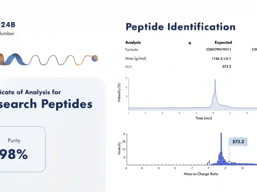
A chemical identifier supports reference and communication. Confirming the identity and characteristics of a physical batch requires suitable analytical methods applied to that batch, with traceable documentation. A web diagram, supplier label, or generic reference spectrum should not be presented as lot-specific verification.
Retatrutide structure FAQs
How many amino-acid residues are in retatrutide?
Curated references describe a 39-residue peptide backbone, including noncanonical residues and a side-chain lipid-linker modification.
What does Aib mean in a retatrutide sequence?
Aib means α-aminoisobutyric acid, a noncanonical amino acid reported at positions 2 and 20 in structure descriptions.
Where is the fatty-diacid group attached?
Structure records describe the C20 diacid-linker appendage as attached to the side chain of Lys17.
Does a chemical structure image verify a sample?
No. A structure image describes a reference molecule. It does not identify the contents of a particular sample or document its batch-specific quality.
References
- Wang L, et al. Structural insights into the triple agonism at GLP-1R, GIPR and GCGR manifested by retatrutide. Cell Discovery. 2024;10:68. doi:10.1038/s41421-024-00700-0.
- National Center for Biotechnology Information. PubChem: Retatrutide. Chemical structure and linked depositor records.
- National Center for Advancing Translational Sciences. Global Substance Registration System: Retatrutide sodium. Substance identity record.
- Jastreboff AM, et al. Triple-Hormone-Receptor Agonist Retatrutide for Obesity—A Phase 2 Trial. New England Journal of Medicine. 2023;389:514–526. doi:10.1056/NEJMoa2301972.
Editorial note: This structural reference summarizes public scientific and chemical records. It is not medical advice, a synthesis guide, or a verification certificate for any physical material. Reviewed October 10, 2026.